Undergraduate Researcher
MISI Group, UCL Hawkes Institute
Jul 2026 — present · London, UK
Funded by the Research Ignition Award, and continuing as my final-year project.

Photo by Denny Müller on Unsplash
The problem
Endoscopic transsphenoidal pituitary surgery has no adequate real-time intraoperative imaging. Optical ultrasound probes built from off-the-shelf fibre are a strong candidate — but reaching the pituitary needs a ≈11.8 mm bend radius, against a manufacturer’s rating of 43 mm short-term and 86 mm long-term for a 400 µm core. Behaviour past that specification was simply uncharacterised.
So: which core diameter gives an anatomy-compatible probe, and what are its safe operating limits?
What I do
Designed the experiment. Six fibre configurations × 13 curvature channels × four actuation speeds — 312 planned runs. Running 400 µm both buffered and unbuffered turns a diameter sweep into a two-factor design, which is the only clean way to separate the coating’s contribution from the core’s.
Built the rig. Laser-cut acrylic clamping a 1.8 mm sheath at thirteen fixed radii, from a straight control to a 90° bend at R = 9.55 mm, on an optical breadboard. Four design iterations, DXF exports, 3D-printed parts, a separately designed camera mount and technical drawings.
Wrote the analysis. Fourteen Python modules, around 3,000 lines: OpenCV marker detection and tracking, SciPy B-spline centreline fitting to convert pixel positions into arc-length along a curving fibre, Savitzky–Golay filtering for velocity, pandas aggregation, a matplotlib/seaborn figure suite. YAML config, argparse CLI, structured logging, pickle caching so a crashed 181-video run resumes rather than restarts, a separate validation harness, and path guards that make it impossible to write into raw source data.
Engineered the quality layer. Calibration of 59.0 px/mm from 1,071 pooled marker separations at 1.6% CV, with the reliability flag and the assumptions recorded. Every statistic gated on a detection rate of 0.6 or better; excluded points drawn as faded open markers rather than silently dropped.

What came out of it
- 200, 300 and 600 µm reach 9.5 mm — inside the 11.8 mm clinical target. 800 µm cannot be driven below ~16 mm and deformed the rig: ruled out for this approach. A clean negative result, and the most decision-relevant thing in the study.
- Tip speed is near-constant across radii at low drive rates (CV ≈ 0.05–0.12).
- The degradation at 5–7 fps is motion blur — an instrumentation artefact, not mechanics. Separating those two was the difference between a false finding and a caveat.
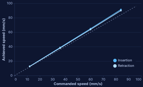
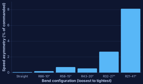
- In the second, mm/s-controlled campaign: 1,715 measured strokes, a monotonic curvature-to-asymmetry relationship rising from 0.06% straight to 8.09% at the tightest radius, and a systematic +7–9% overshoot between commanded and achieved speed — so the commanded value cannot be used as ground truth.